PUSHING THE LIMITS OF DIGITAL: Accuracy of Straumann EXACT™ vs. the Analog Gold Standard – A Case Report.
Objectives
The primary aim of this case
report
is to evaluate the fully digital Straumann EXACT™ workflow
against the traditional analog laboratory workflow.
Can a fully digital workflow
truly match the accuracy of the analog gold standard?
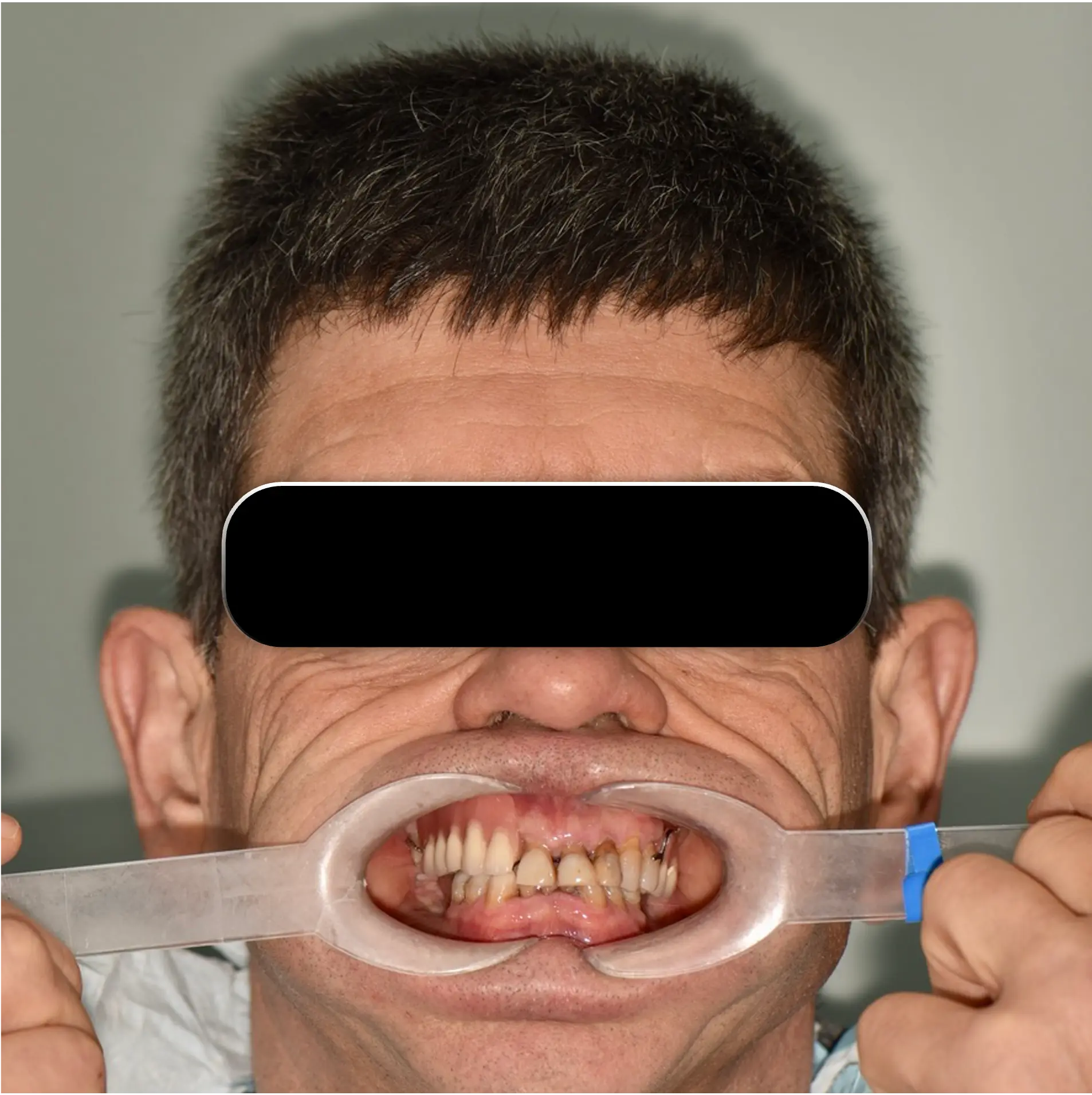
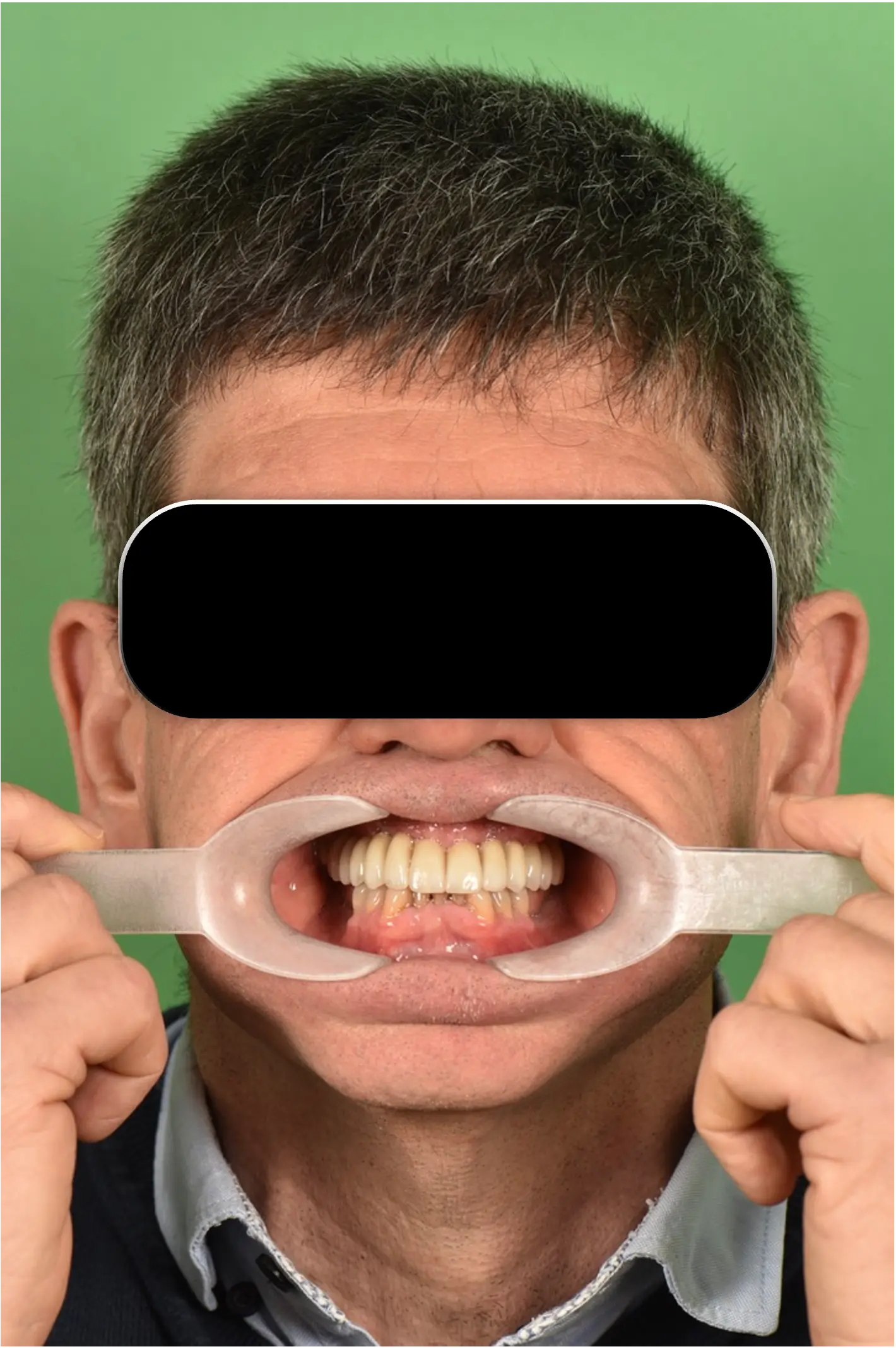
Initial Presentation
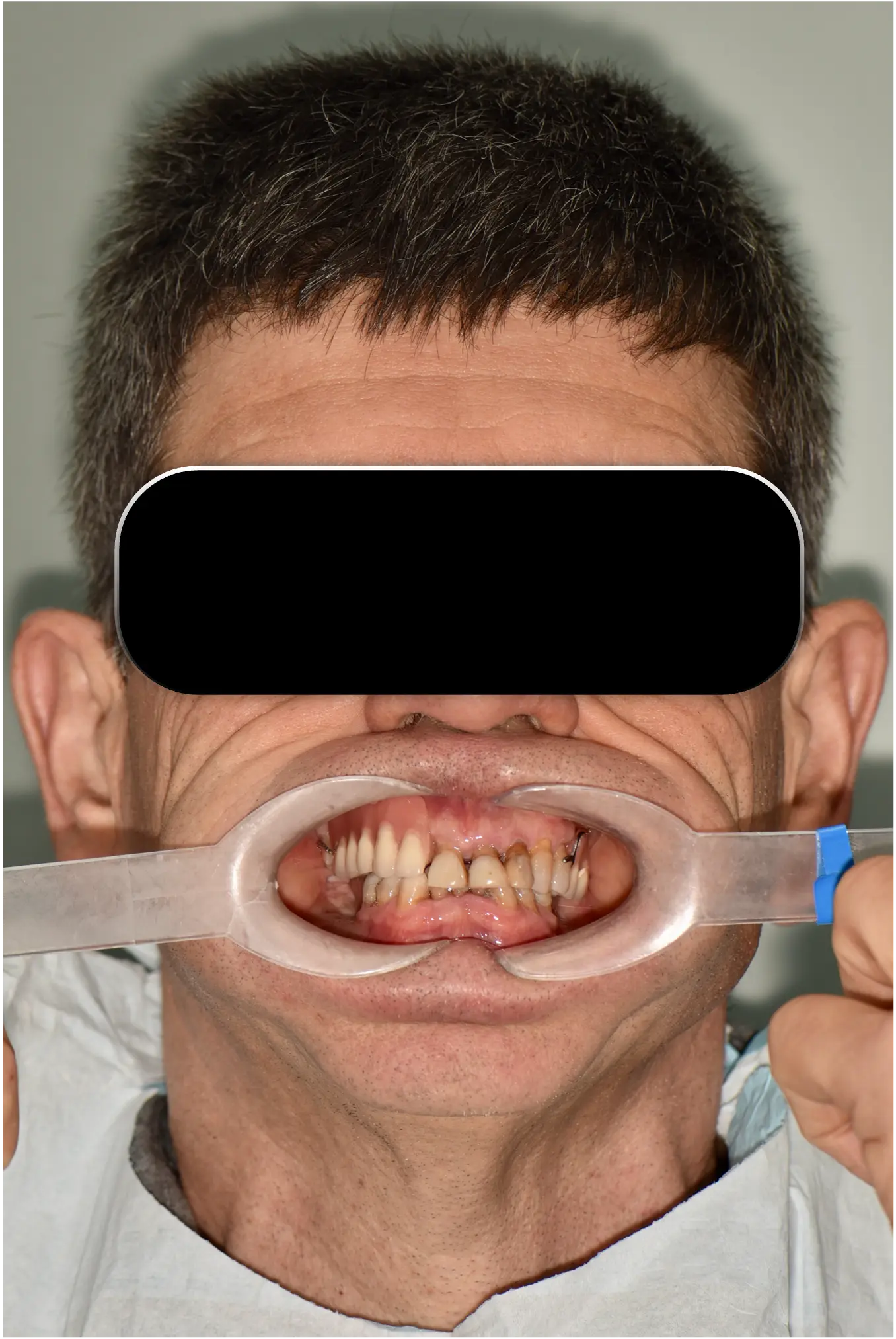
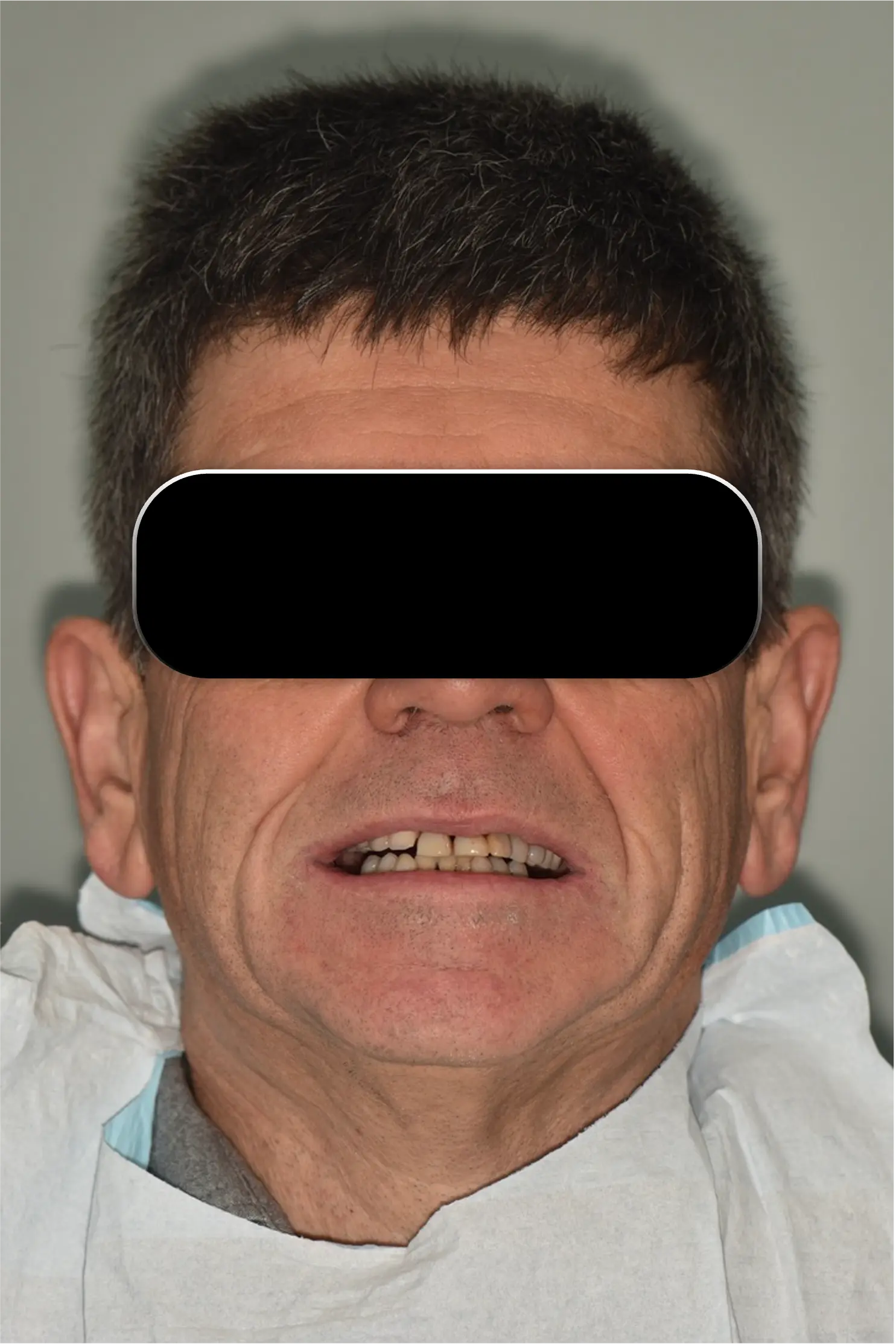
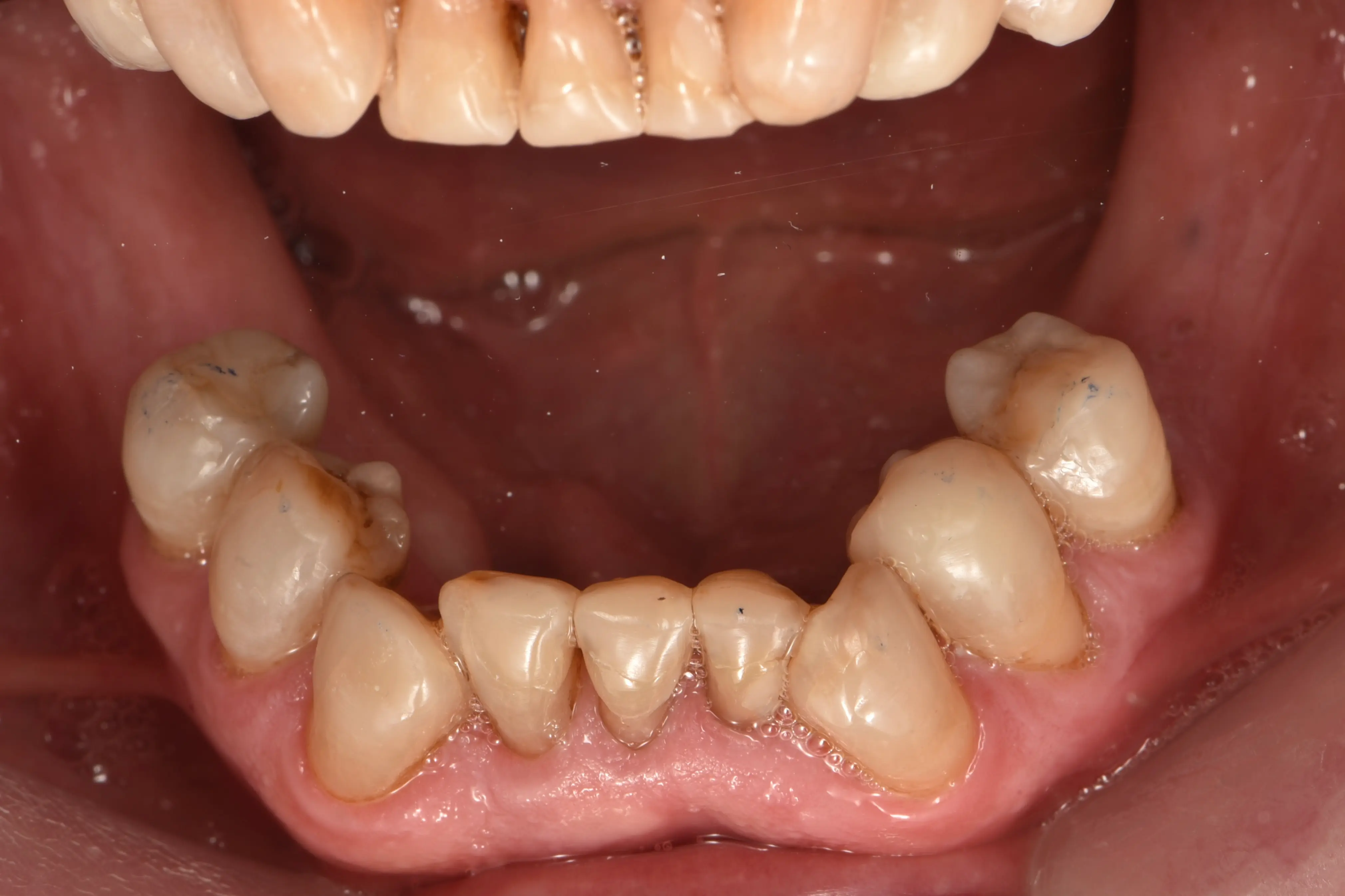
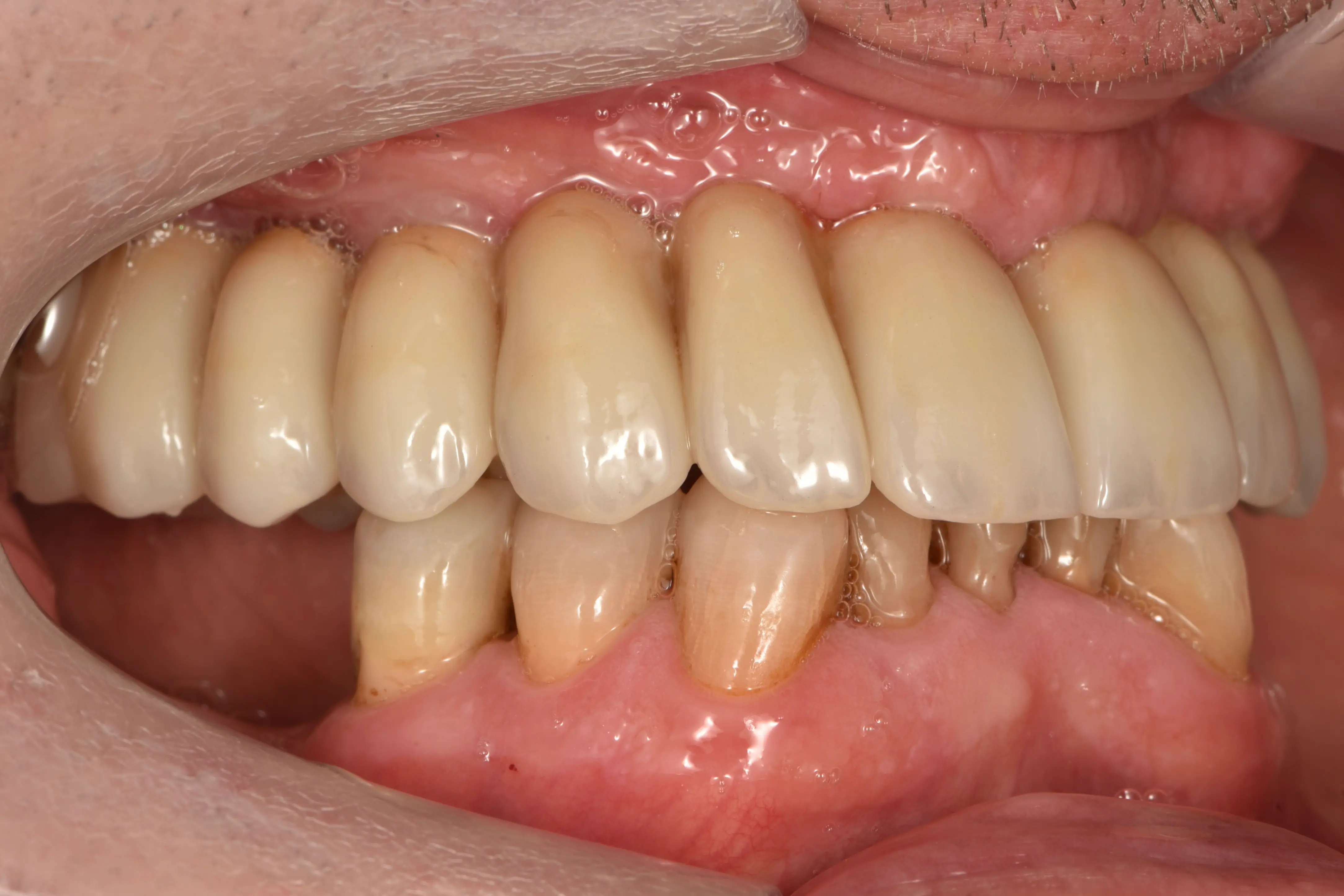
- A 60-year-old male patient presented with a maxillary partial denture, a mandibular shortened dental arch, and severe tooth wear.
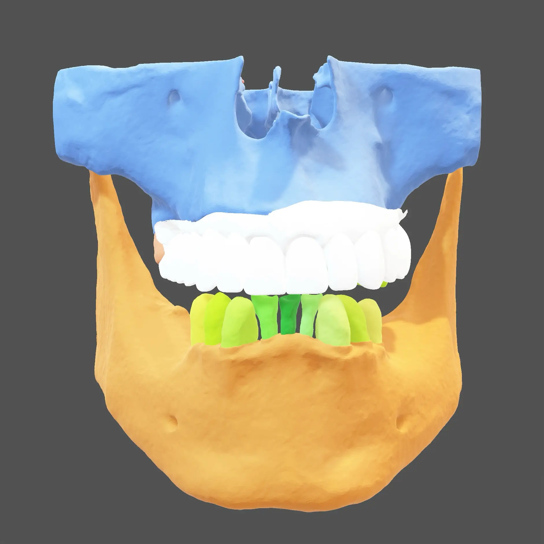
Digital Wax-Up
- A digital wax-up guided the treatment plan.
- The mandibular tooth wear was restored first to establish a correct vertical dimension.
Surgery & Implantation
- A base, surgical, and prosthetic guide, along with a provisional bridge, were fabricated via the Straumann Smile in a Box platform.
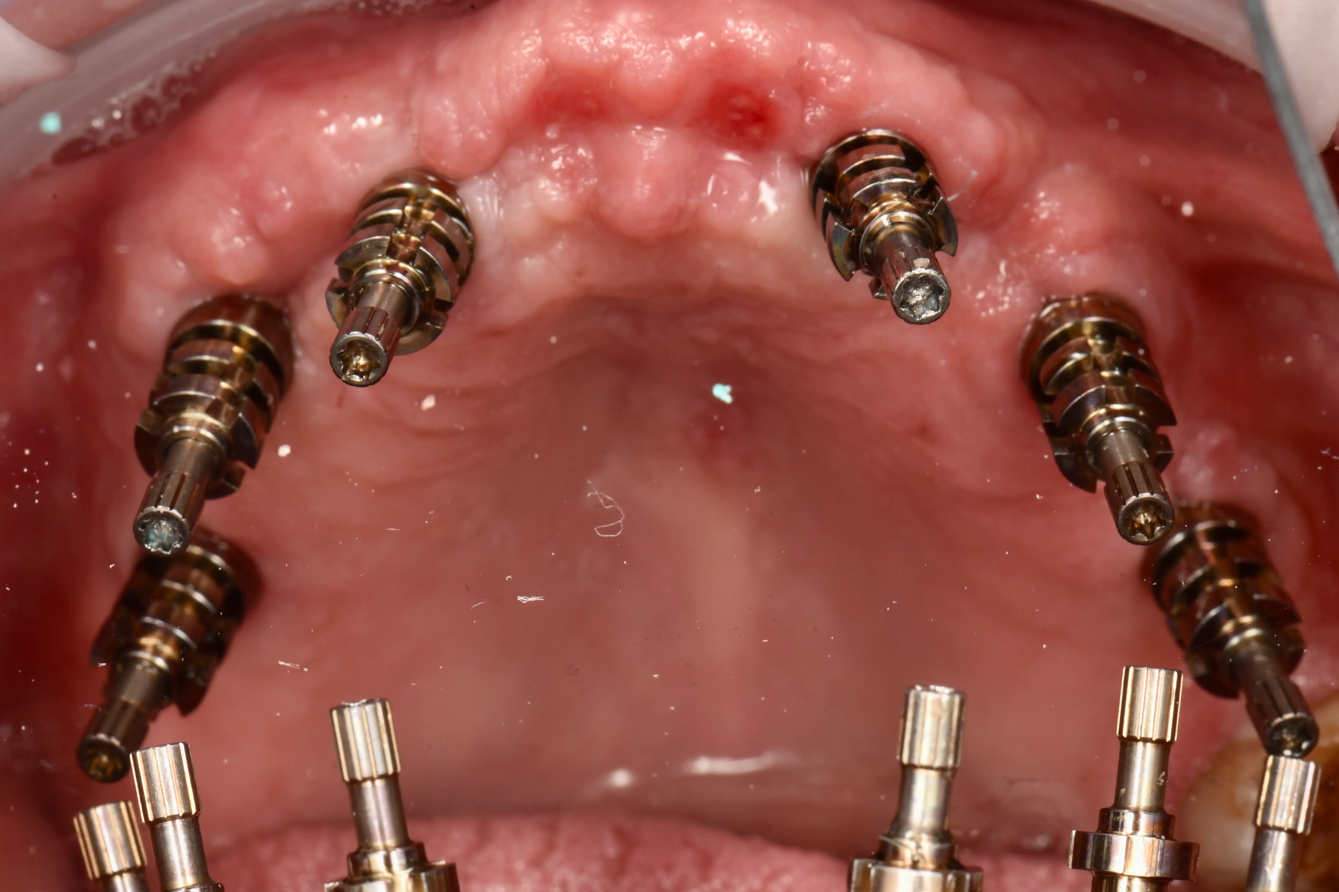
- The maxillary teeth were extracted, and six Straumann BLX implants were placed.
- Screw-retained abutments and temporary cylinders were seated.
Intra-Oral Pick-Up
- The provisional bridge was placed using the prosthetic guide.
- It was intra-orally picked up using composite to secure the correct position.
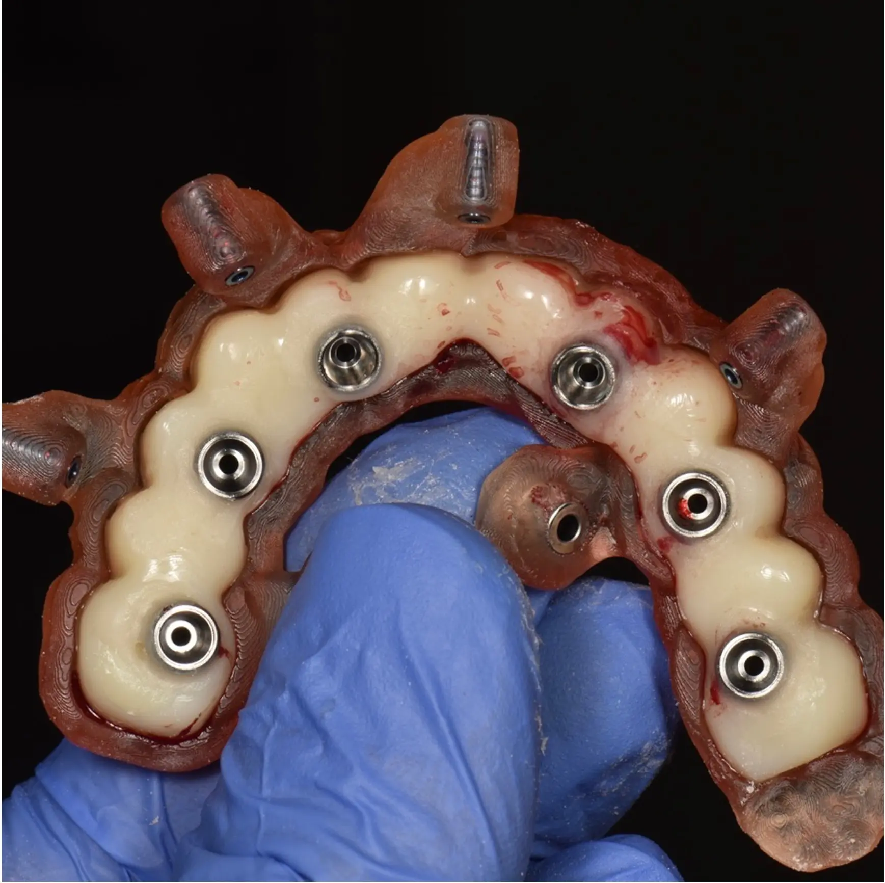
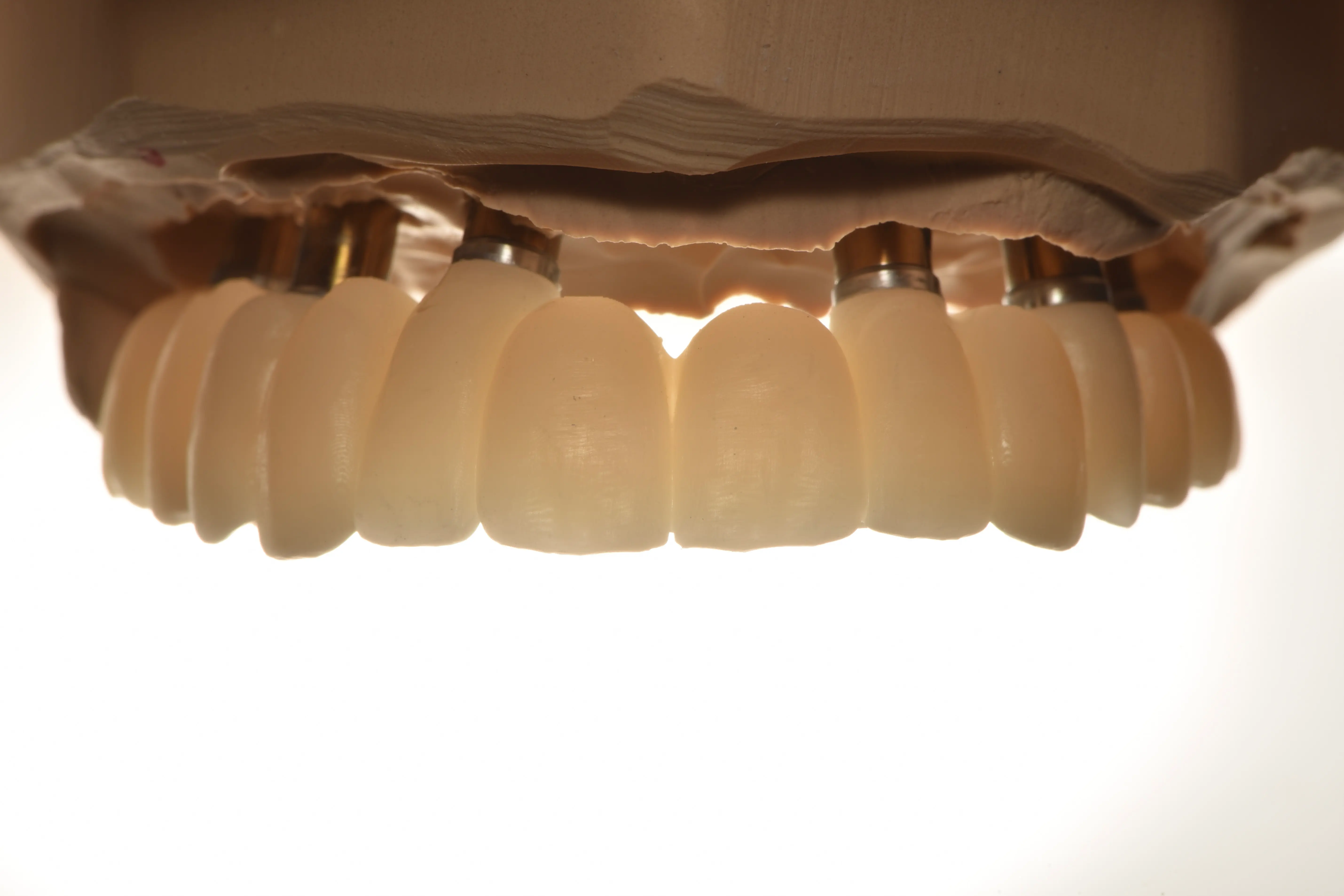
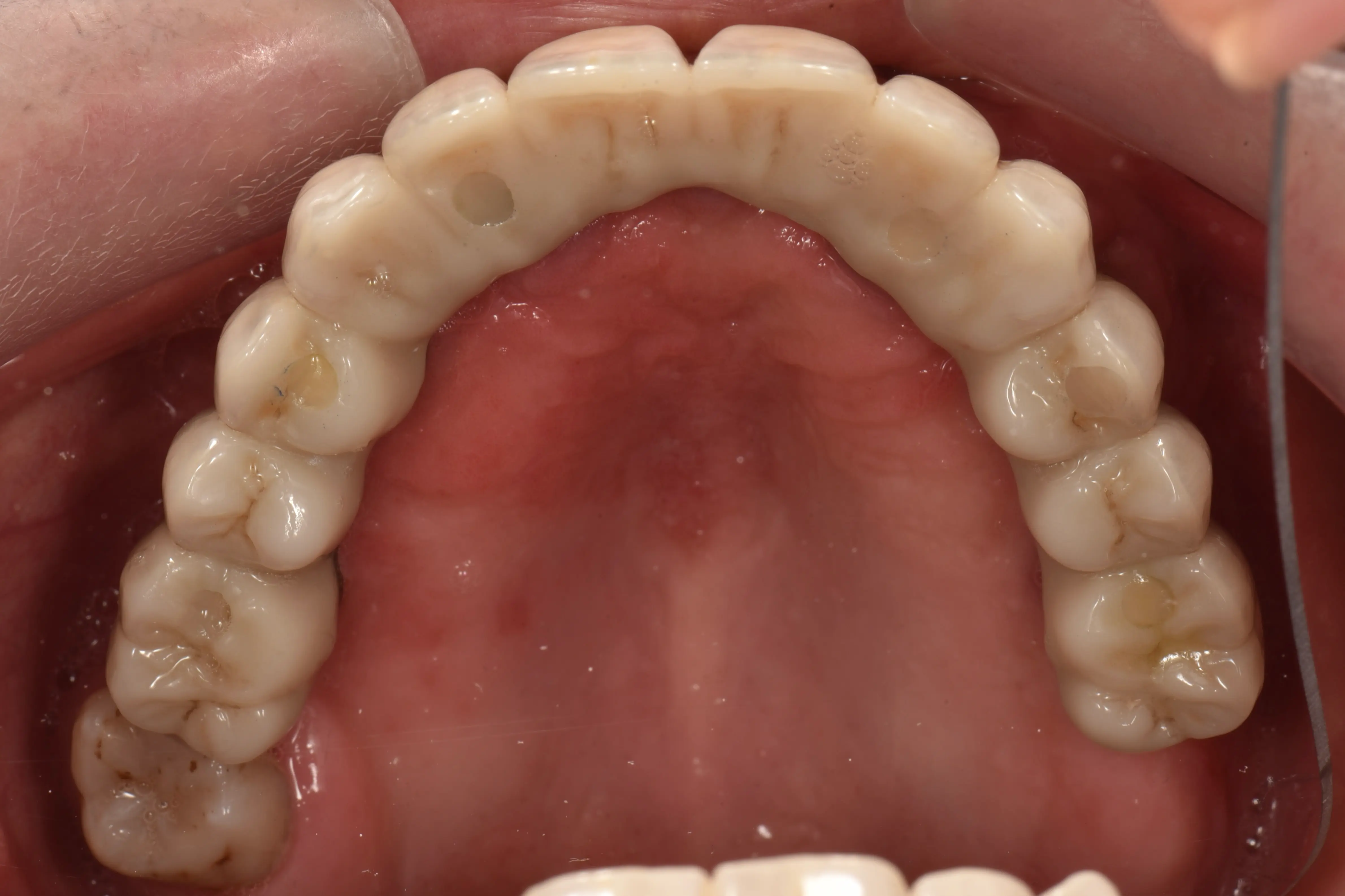
Final Delivery
- The splinted bridge was carefully removed.
- It was extra-orally finished with Unifast III.
- Finally, the bridge was re-inserted into the patient's mouth for final delivery.
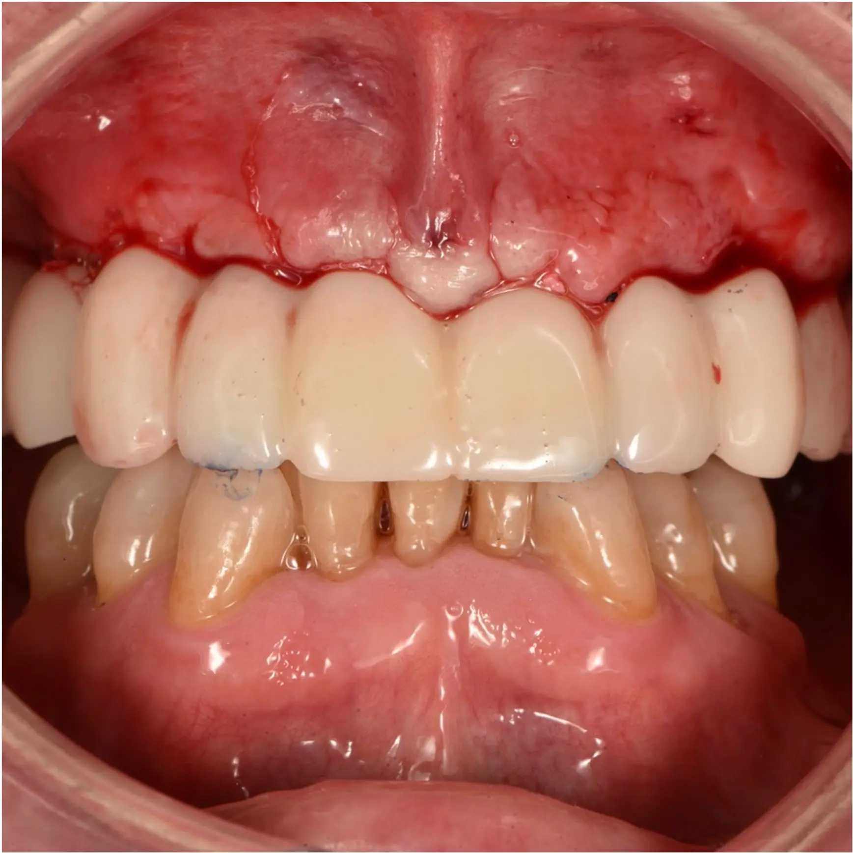
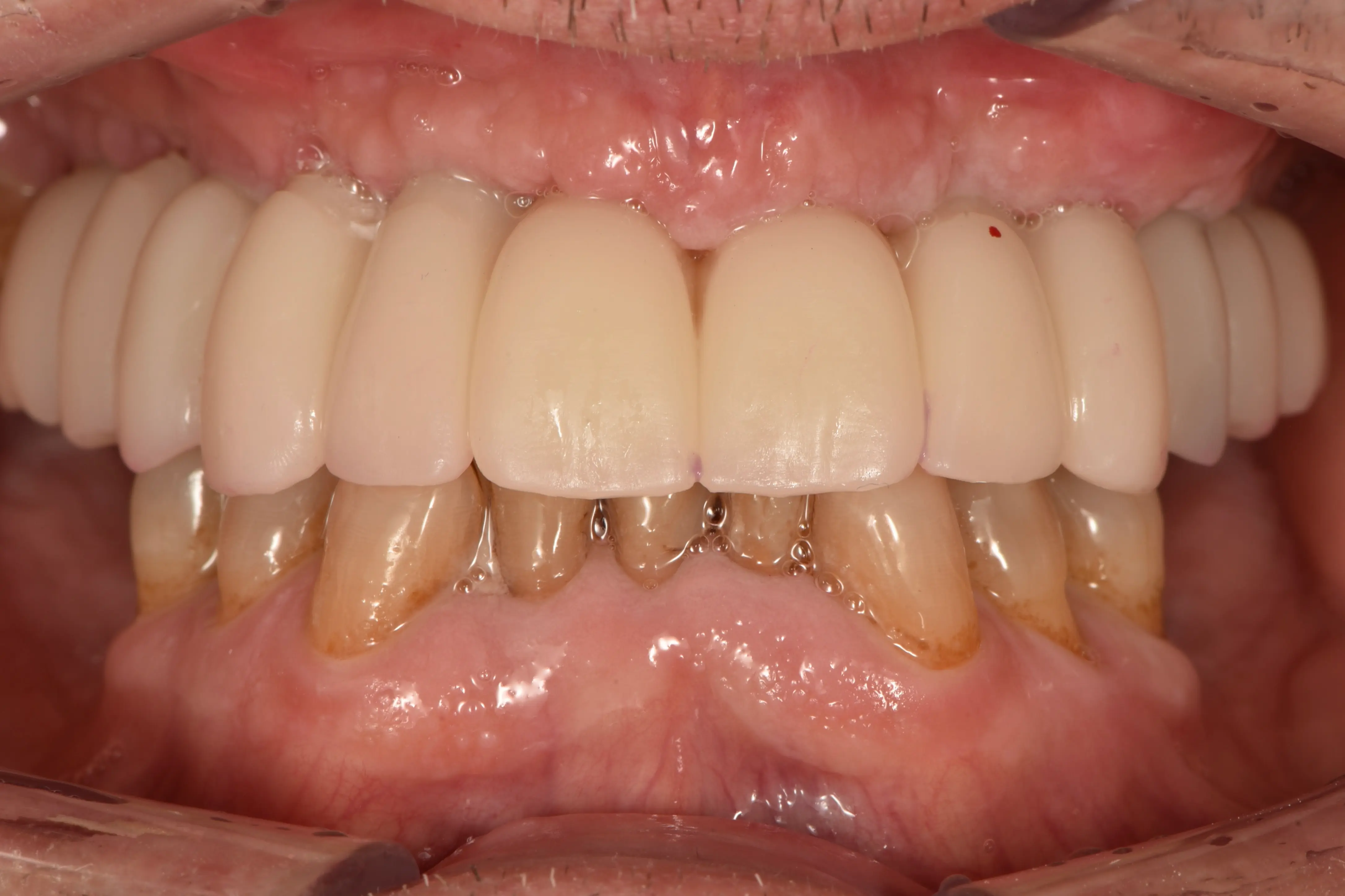
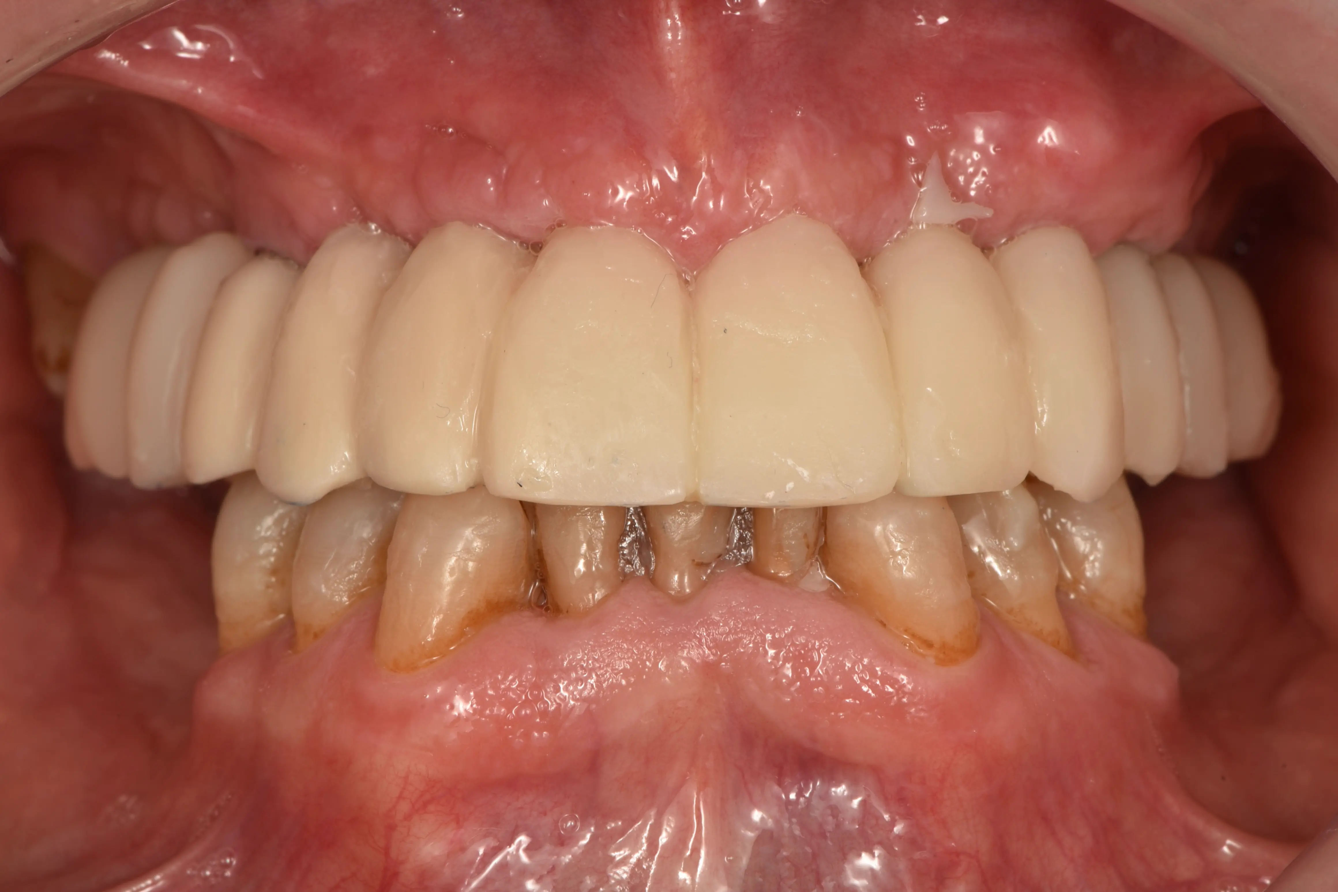
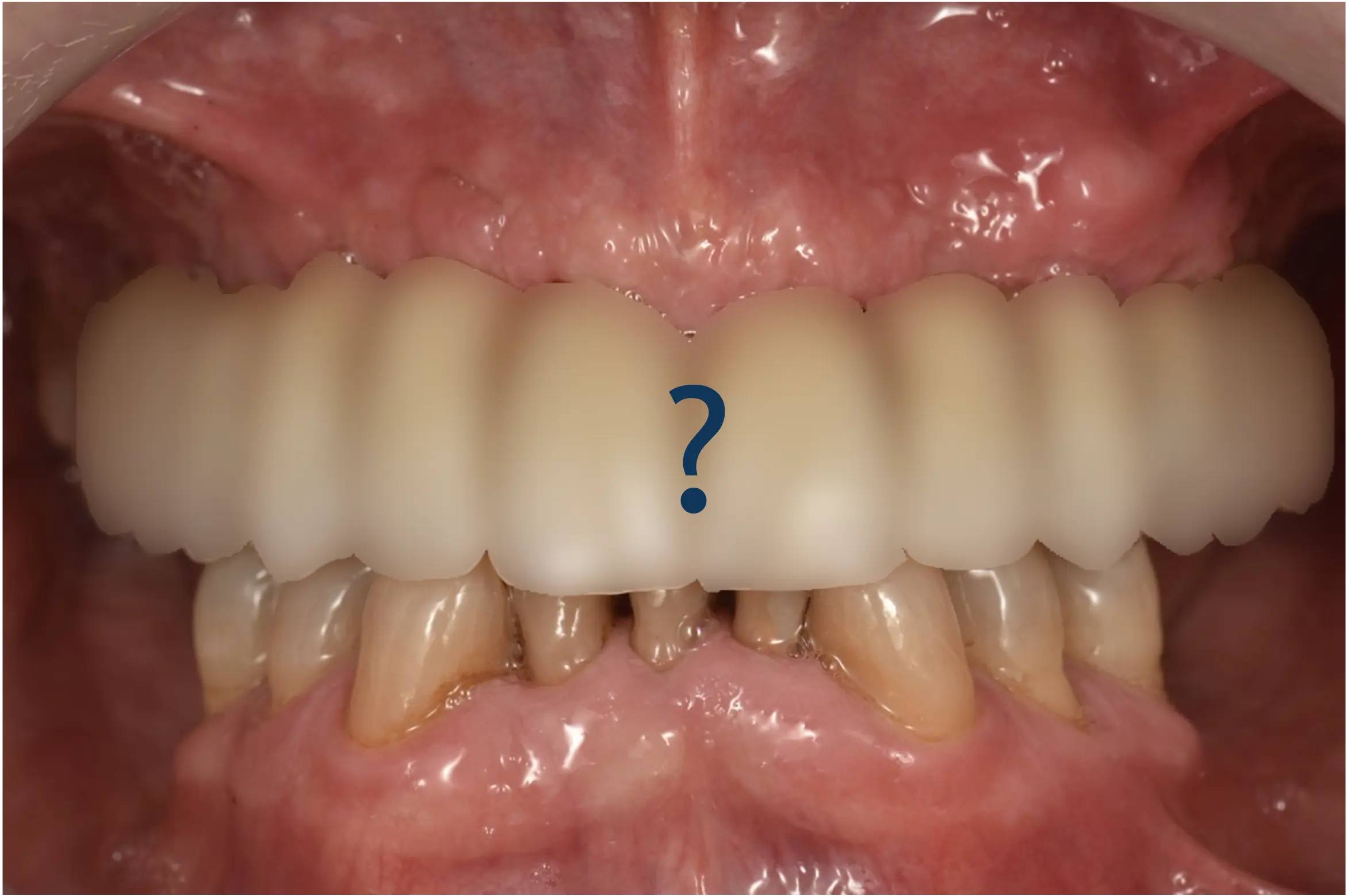
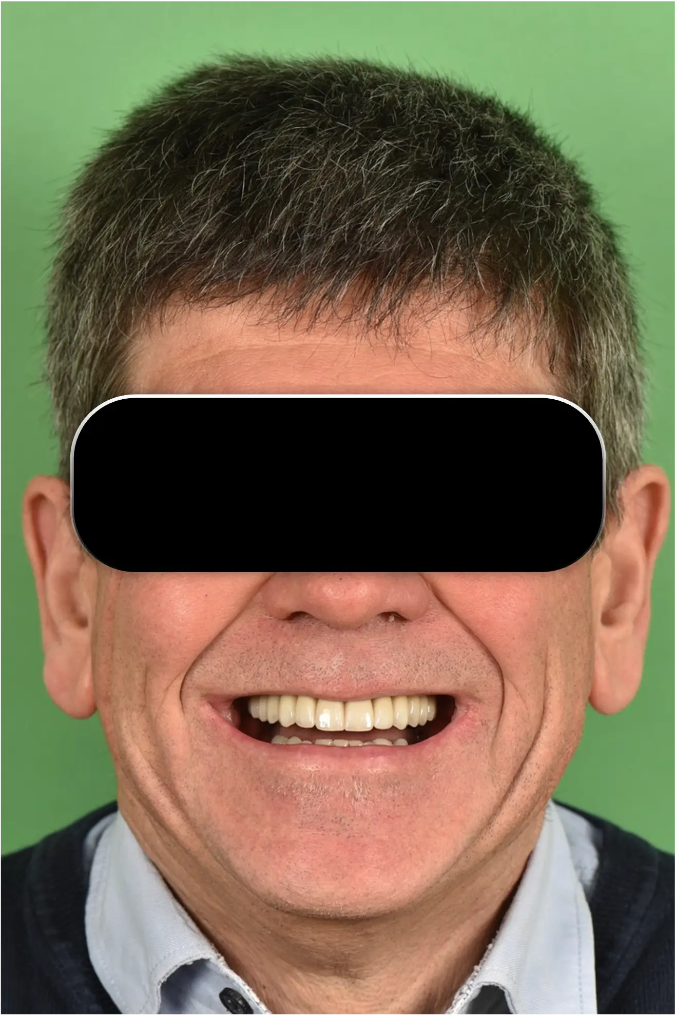
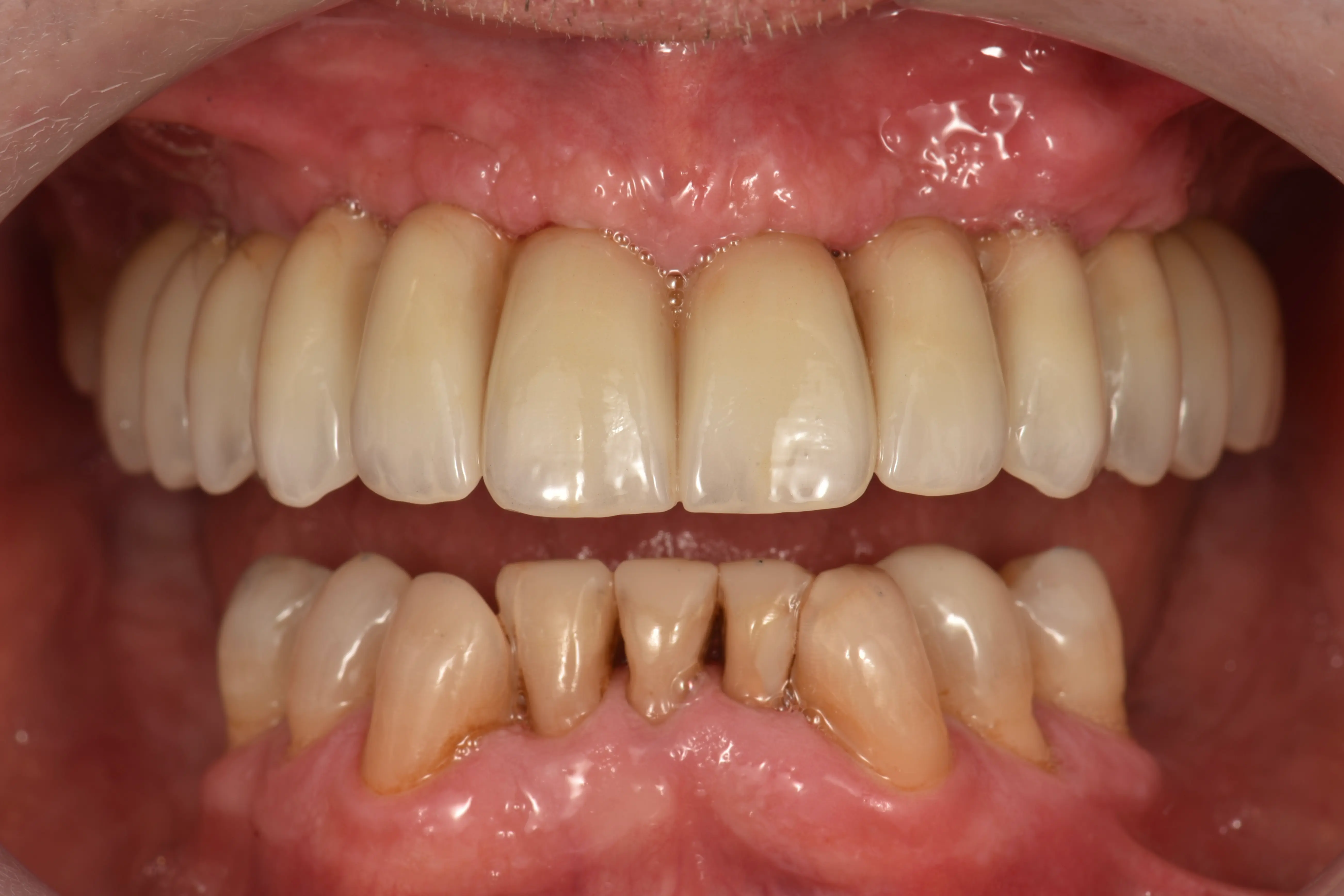
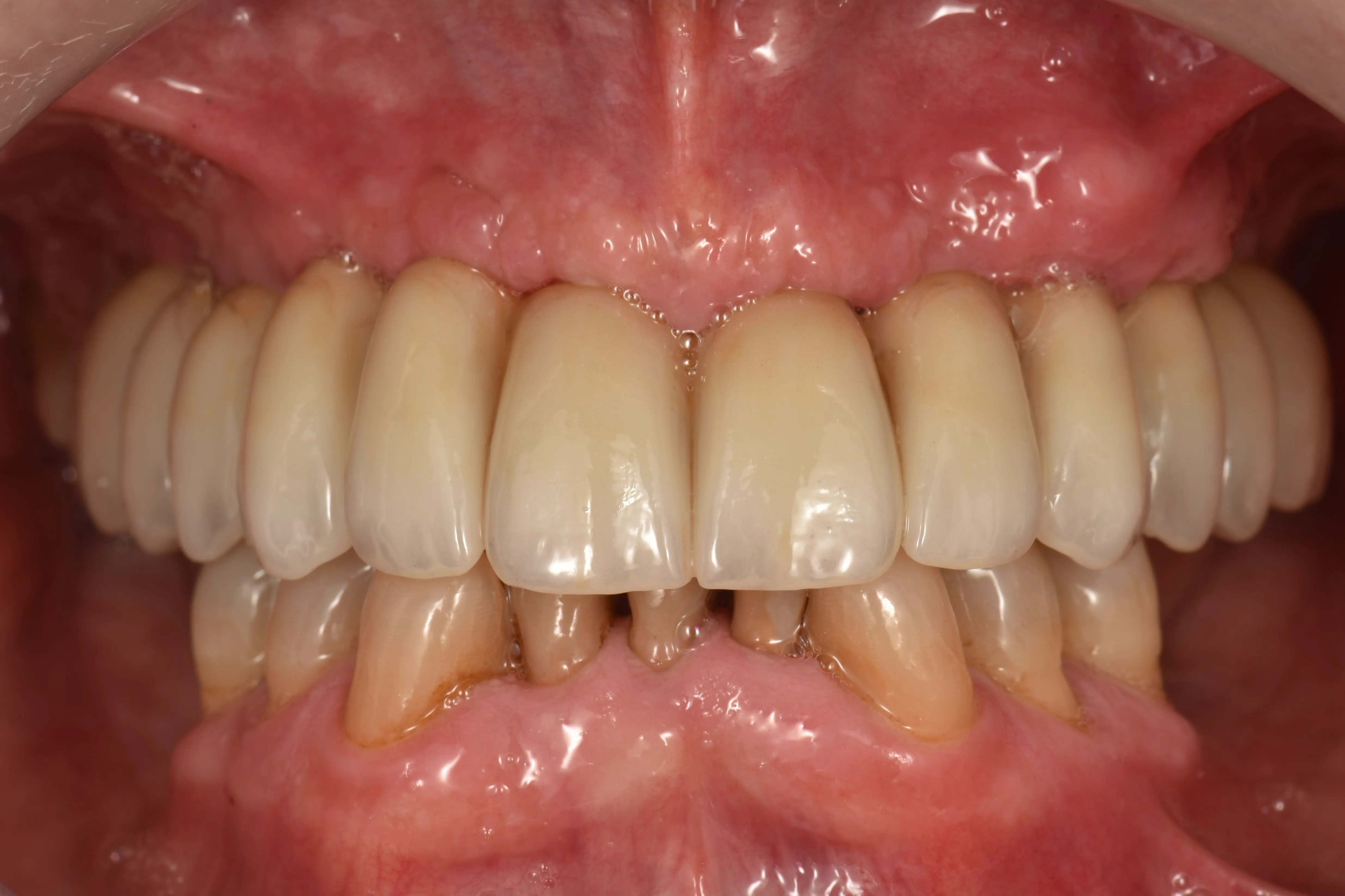
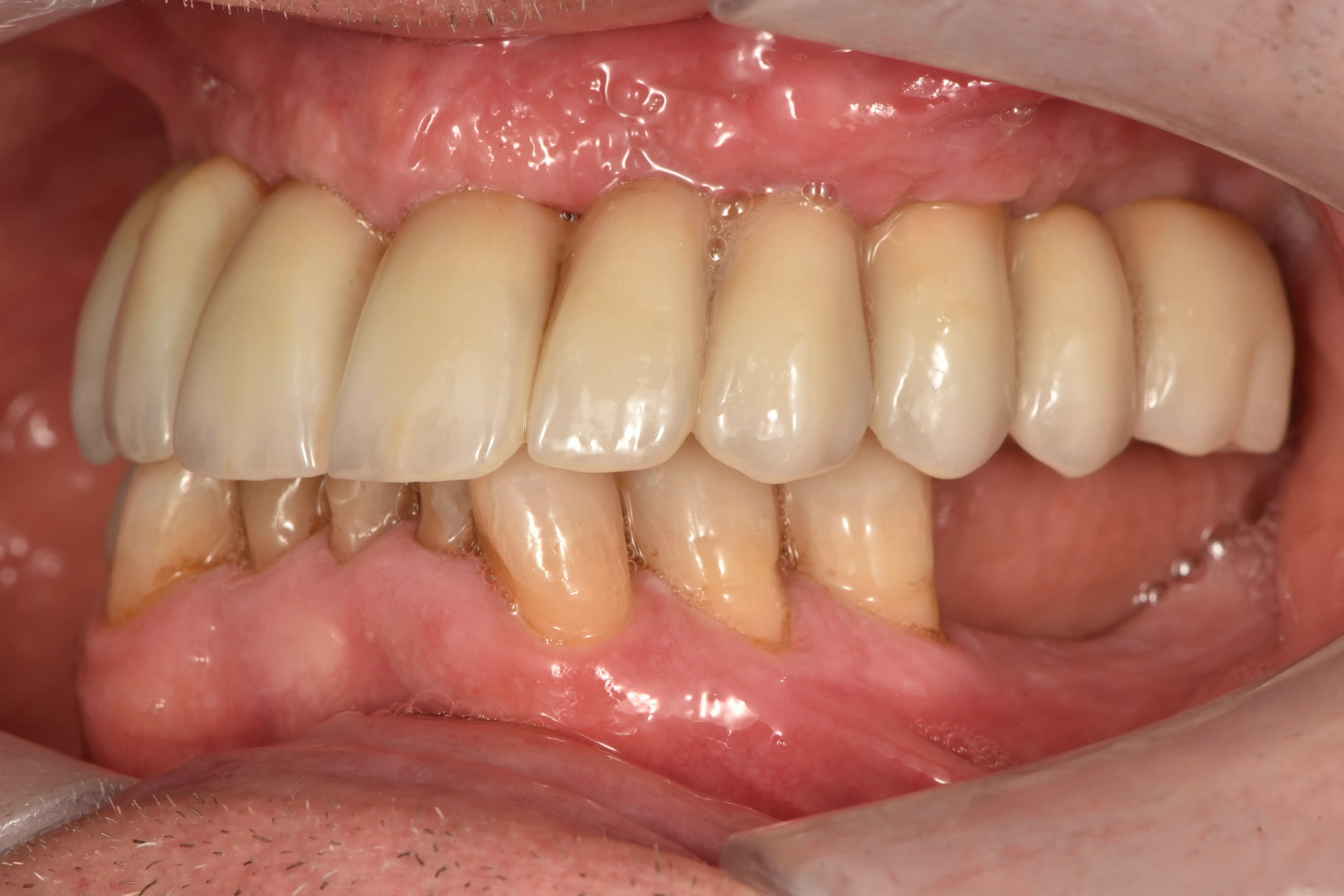
Restoring Function & Aesthetics
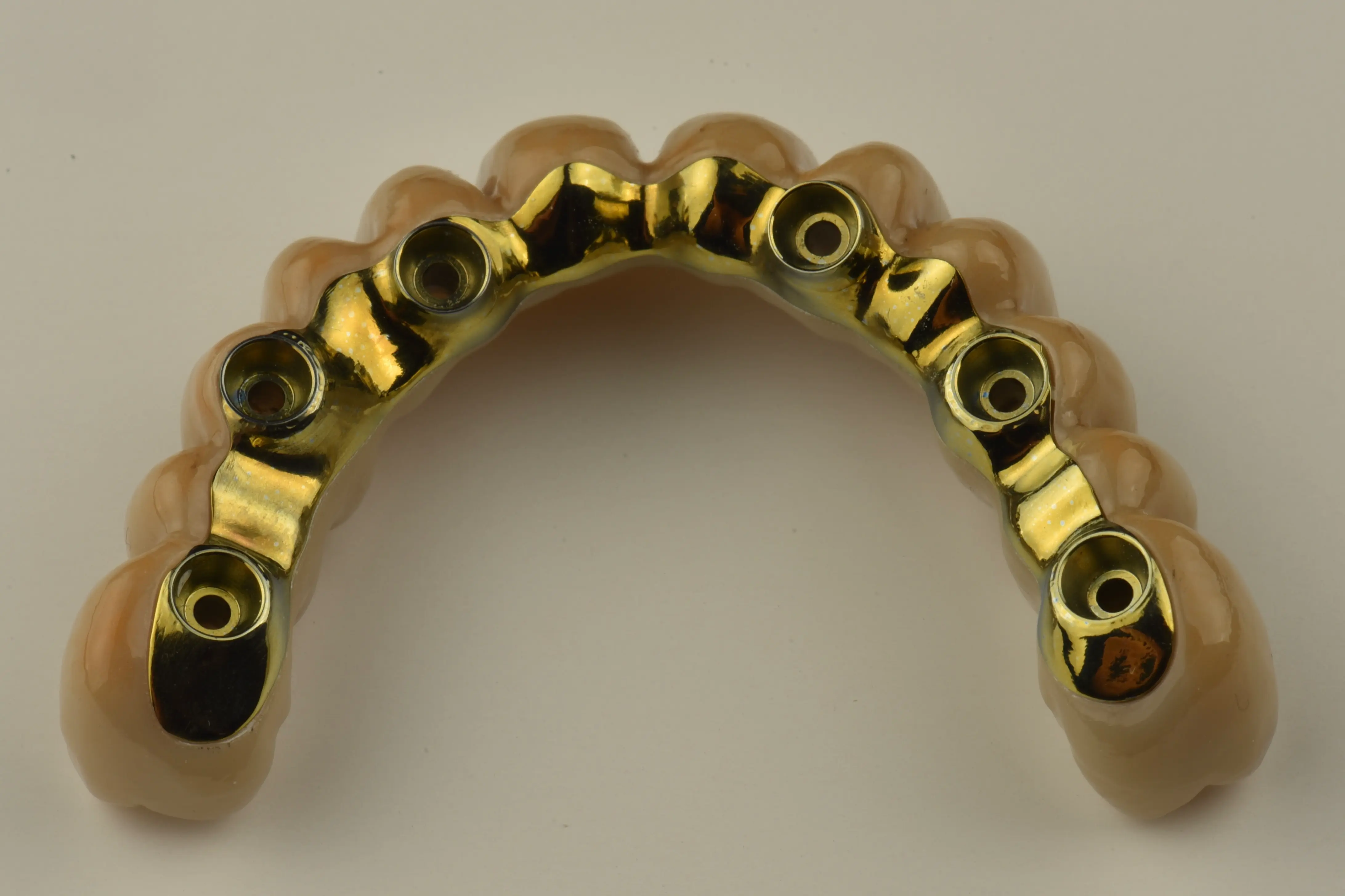
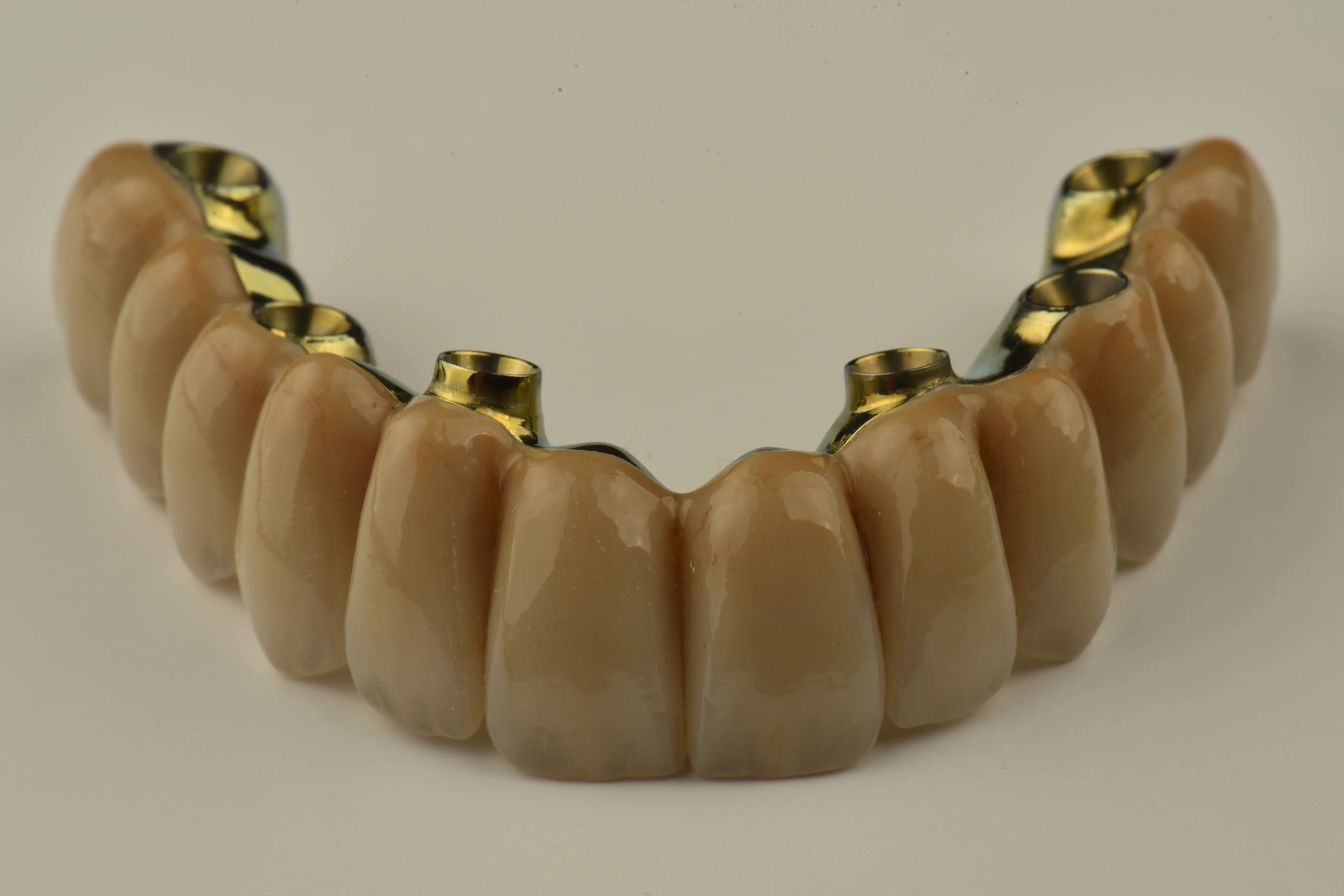
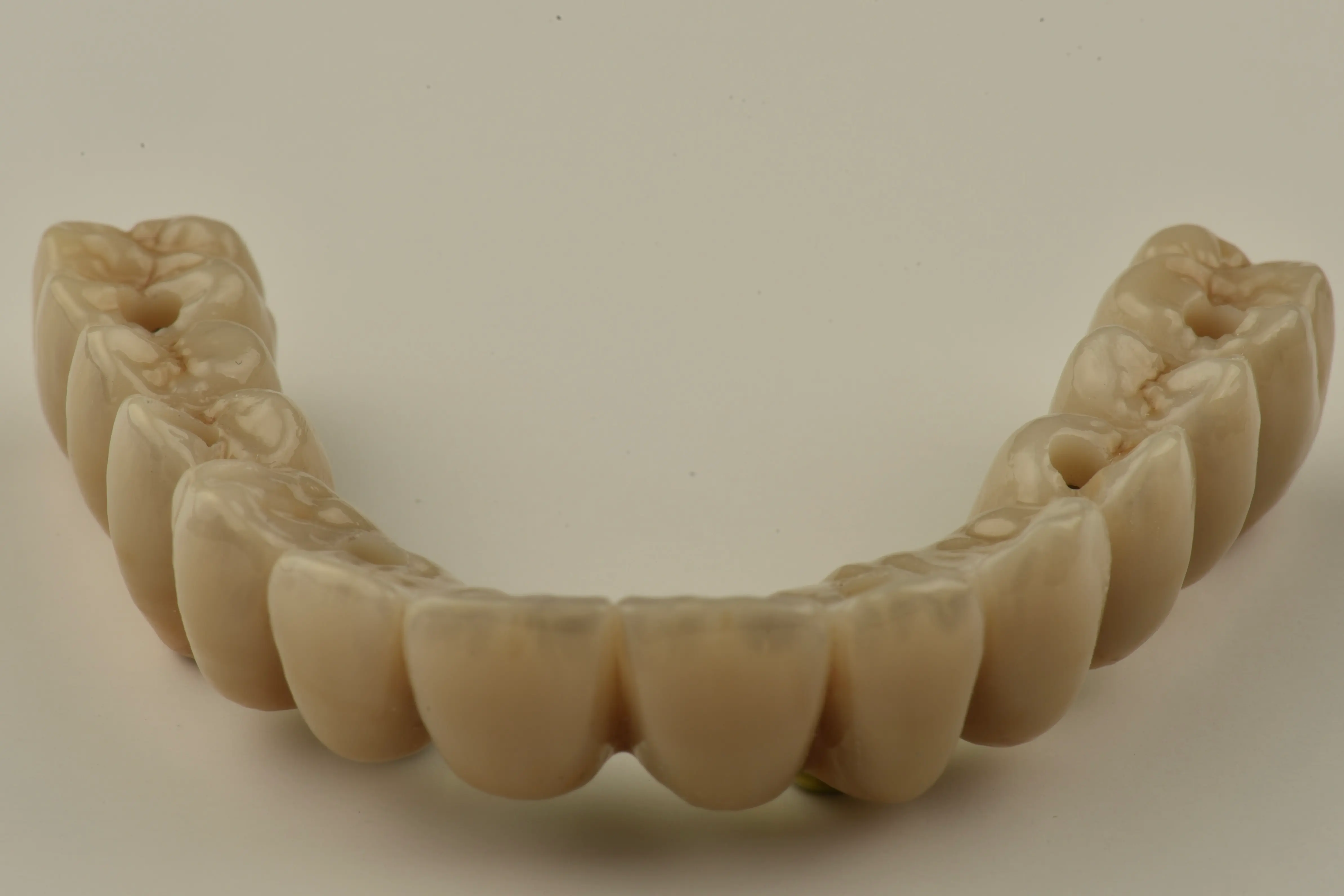
- A definitive Fixed Prosthesis 1 (FP1) bridge restores oral function, aesthetics, and quality of life.
- A digital wax-up guided the Vertical Dimension of Occlusion (VDO), facial proportions, and restorative space.

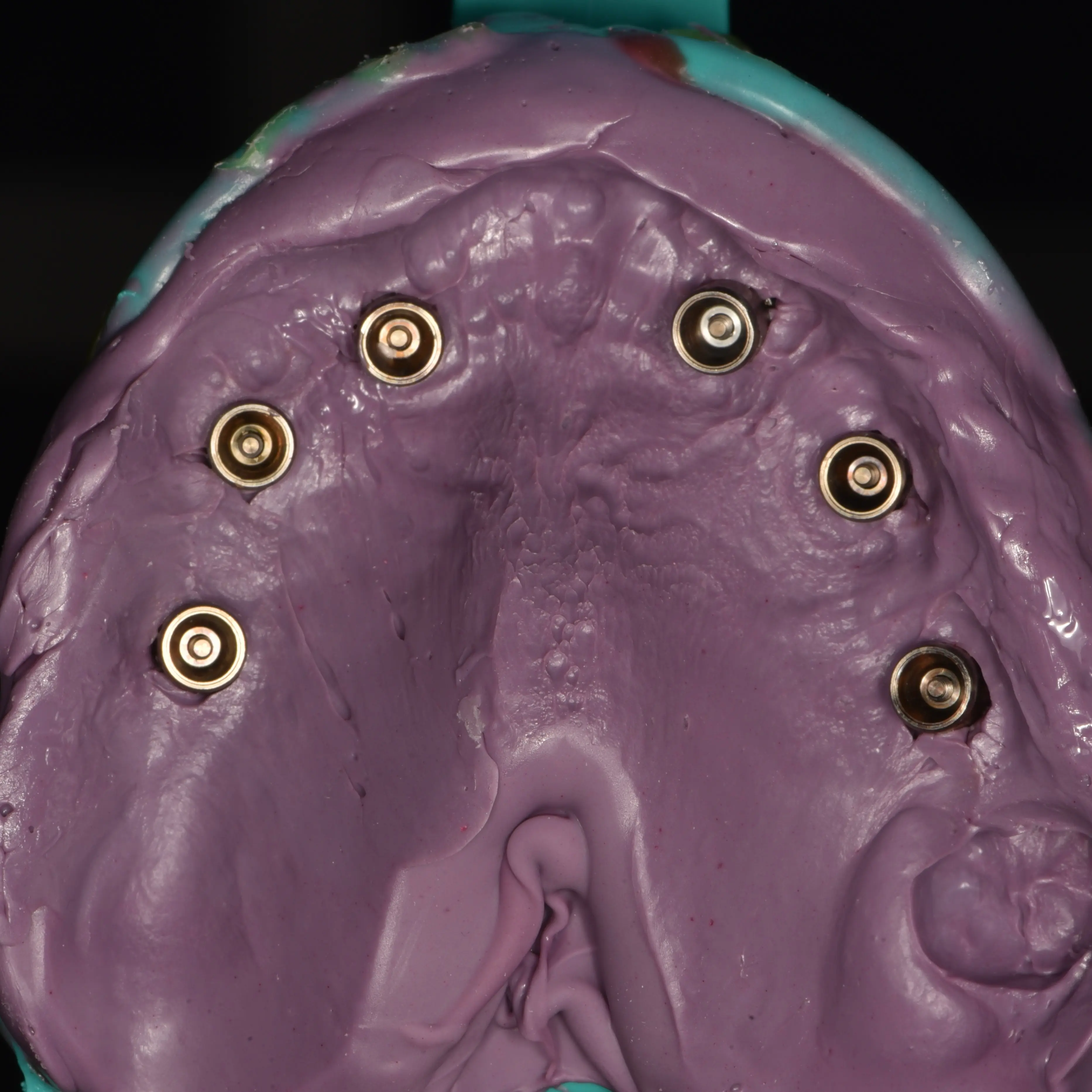
Protocol Evaluation
- To evaluate the EXACT™ protocol, an Impregum impression was taken as the analog reference.
- Utilizing 'Smile in a Box' and Simeda milling kept the entire workflow seamlessly within the Straumann ecosystem.

Straumann EXACT™ Workflow
Choose Treatment Protocol
Select a method to view the clinical workflow
Straumann EXACT™
Fully Digital Workflow
Analog Impregum
Conventional Workflow
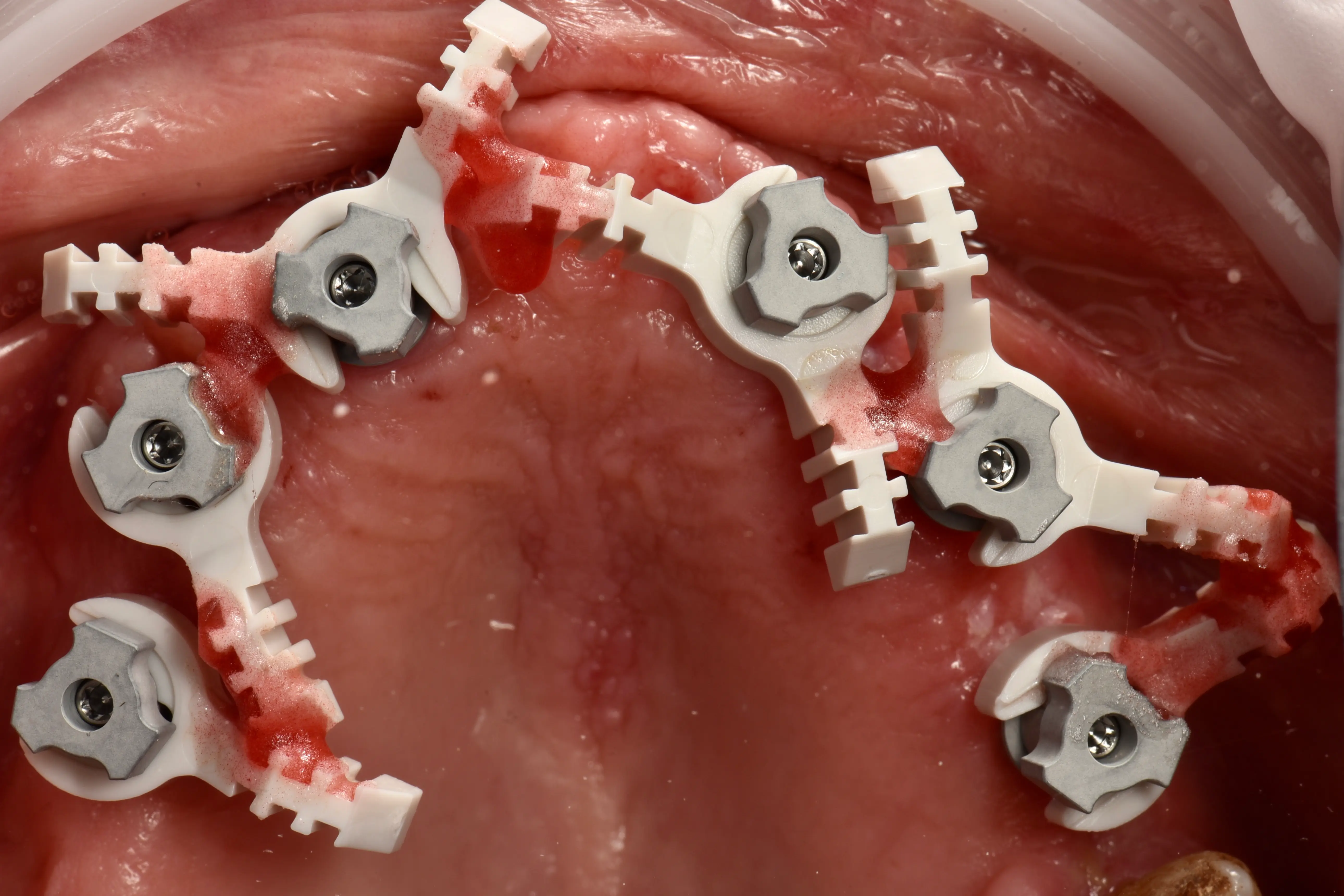
Straumann EXACT™ Scan Protocol
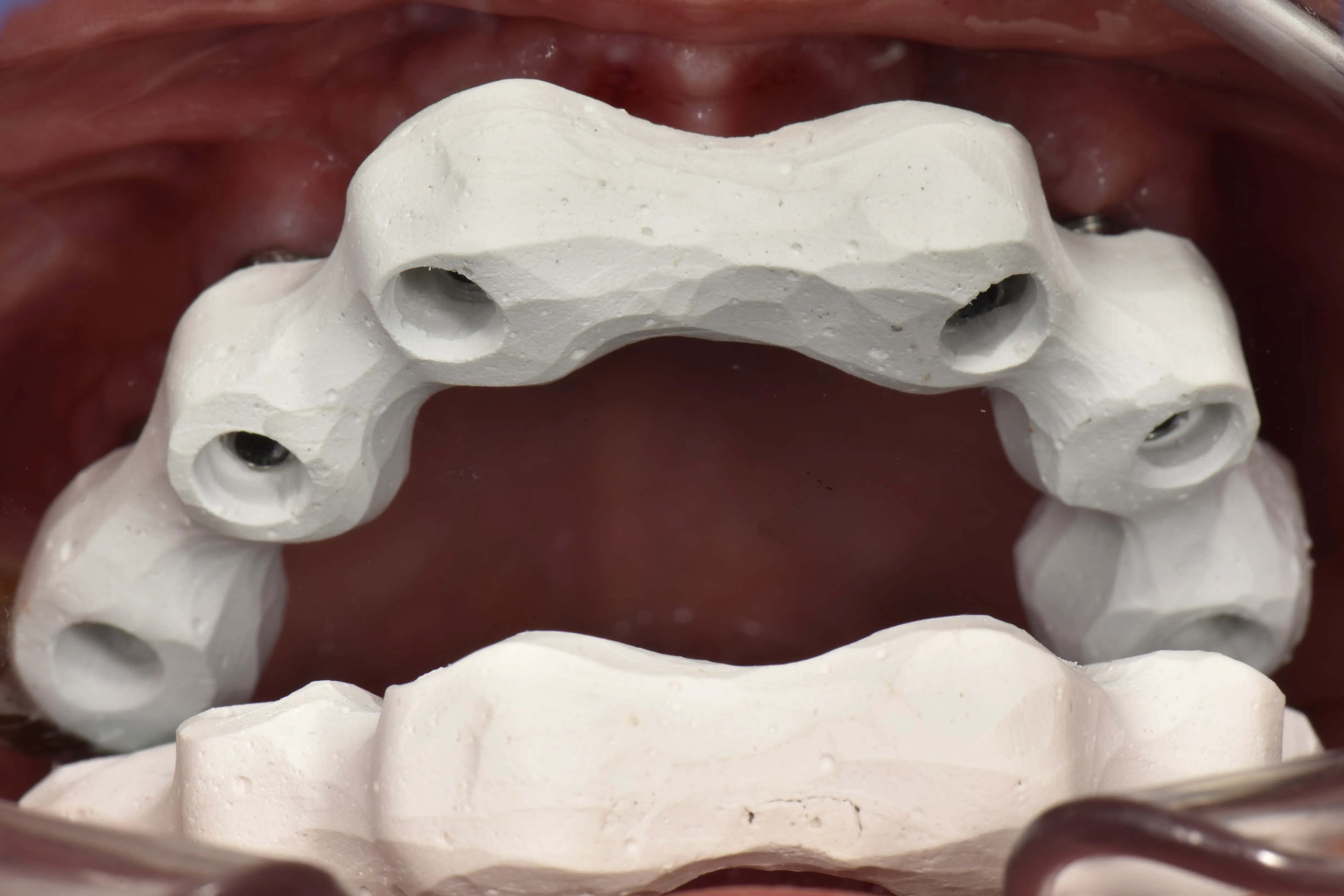
- Splinting Technique (Straumann EXACT™ protocol): 'BASE' scanbodies and 'LINKS' were rigidly splinted with pattern resin to overcome traditional full-arch IOS stitching limitations.
- Maxillary Scans: Digital acquisition included a pre-preparation scan of the temporary bridge, a scan of the edentulous maxilla, and a scan of the splinted scanbodies.
- Mandibular Scan: A scan of the opposing dentate mandible.
- Photography: Standardized clinical documentation.
Provisionalization
- Placement of a provisional bridge (torqued to 15 N/cm), digitally designed using clinical photos, lab communication, and the existing prosthesis.
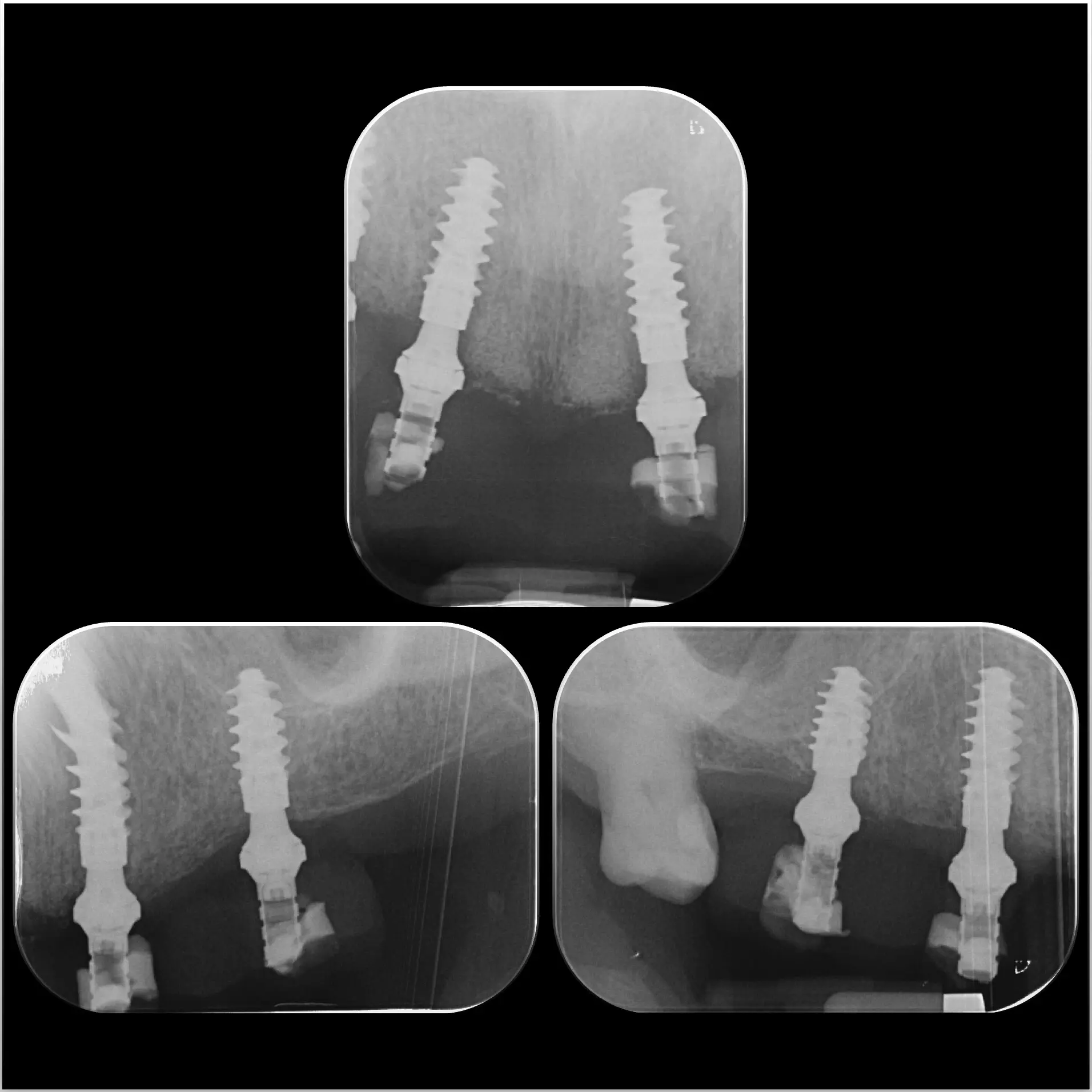
- Three periapical radiographs taken for baseline control.
Clinical evaluation & refinement
- Evaluation of the provisional after one month of functional loading.
- Modifications to occlusal plane, incisal length, buccal contours, and pontics.
- Refinements communicated to the lab to finalize the definitive design.
Final delivery
- Definitive bridge fabricated on the analog model due to a 0.66° (see results) digital deviation.
- Radiographic verification, 15 N/cm torque, and screw channels sealed.
- Follow-up: One-month check-up confirmed stable occlusion and oral hygiene.
Three Digital Models
- Model 1 (Analog Workflow): Lab scan of the Impregum-derived master stone model.
- Model 2 (Direct Digital): Intra-oral Straumann EXACT™ scan.
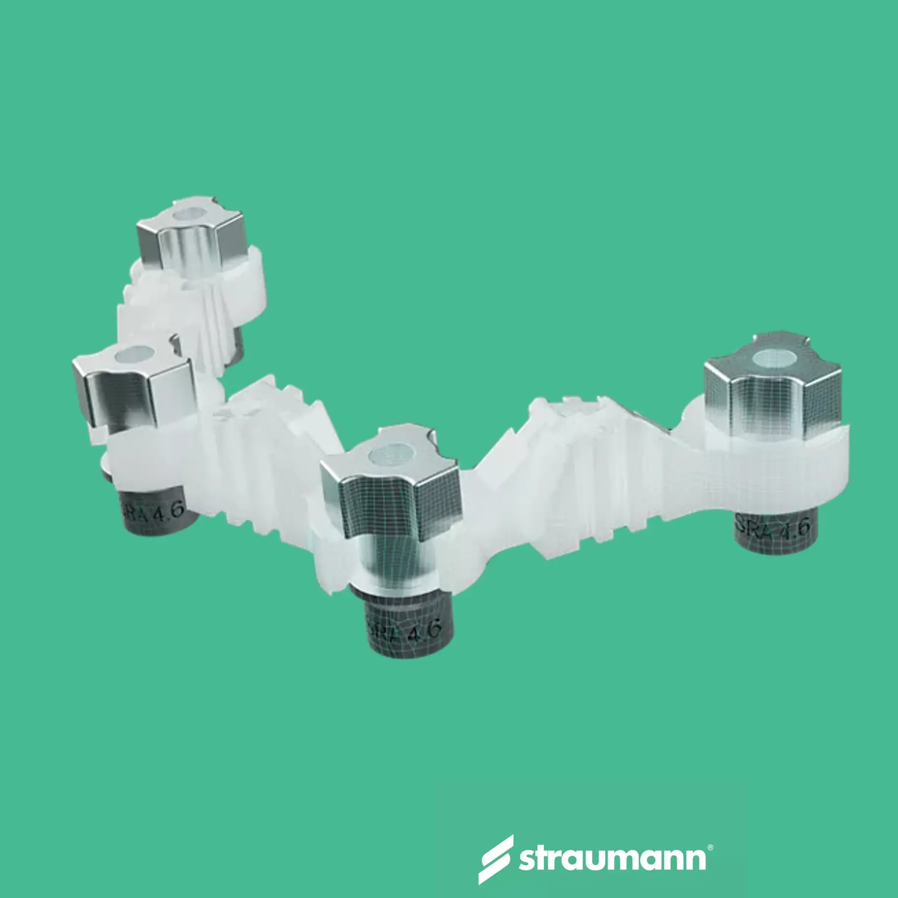
- Model 3 (Hybrid Digital): Extra-oral lab scan of the splinted EXACT™ structure using RevEX™ scanbodies.
Model 1 vs Model 2 (EXACT™)
- The implant positions were converted to 6 virtual cylinders (4.5 x 7 mm) to assess precision.
- Part Comparison: Evaluating 3D spatial deviations between the analog cast and the direct digital scan.
- Angulation Comparison: Measuring the exact angular differences in degrees.
Model 1 vs Model 3 (Hybrid)
- The same volumetric and angular comparisons were executed for the extra-oral scan using RevEX™ scanbodies.
- This highlights the differences between a direct intra-oral scan and an extra-oral splinted approach.
Deviation Analysis Summary
| Part comparison | Mean (mm) | Median (mm) | RMS (mm) | Max Diff (mm) | SD (mm) |
|---|---|---|---|---|---|
| Analogue vs. Straumann EXACT™ | 0.0409 | 0.0375 | 0.0490 | 0.1509 | 0.0271 |
| Analogue vs. Straumann RevEX™ | 0.0679 | 0.0551 | 0.0875 | 0.2358 | 0.0552 |
| Angular Deviation | Tooth 16 | Tooth 14 | Tooth 12 | Tooth 22 | Tooth 24 | Tooth 26 |
|---|---|---|---|---|---|---|
| Analogue vs. Straumann EXACT™ | 0.66° | 0.48° | 0.11° | 0.13° | 0.26° | 0.49° |
| Analogue vs. Straumann RevEX™ | 0.44° | 0.67° | 0.83° | 0.16° | 0.37° | 0.68° |
Clinical Conclusions
- Workflow Validation: The Straumann EXACT™ protocol demonstrates superior dimensional trueness (0.0409 mm) compared to the hybrid RevEX™ approach.
- Safeguard Protocol: While EXACT™ significantly minimizes errors, peak deviations confirm that analog cast finalization remains the gold standard for full-arch rehabilitations.
Reference: Zhang, Y., et al. (2021). Accuracy of full-arch digital implant impressions: A systematic review. PubMed, 14(2), 157–179.
View Full Article (PDF)Department of Restorative Dentistry, KU Leuven • In collaboration with Labo Hoet.